
Retained Intraocular Foreign Bodies
The reaction of the eye to a retained foreign body varies greatly depending on the foreign body’s chemical composition, sterility, and location. Inert, sterile foreign bodies such as stone, sand, glass, porcelain, plastic, and cilia are generally well tolerated. If found several days after the injury, they may be left in place if they are not obstructing vision. Evaluation for retinal toxicity with electroretinography may be helpful in some cases.
Common reactive foreign bodies are zinc, aluminum, copper, and iron. Zinc and aluminum tend to cause minimal inflammation and may become encapsulated. If very large, however, any foreign body may incite inflammation, causing glial and/or fibrovascular proliferation into the vitreous preretinally and along the ciliary processes. Tractional retinal distortion and detachment and phthisis bulbi may result in loss of useful vision. Migration of the foreign body also can occur, especially with copper.
Pure copper (eg, wire, percussion cap) is especially toxic, and prompt removal is required. Pure copper causes acute chalcosis with severe inflammation and may lead to loss of the eye. Late removal may not cure the chalcosis, which has been reported to increase after surgery in some cases because of dissemination of the metal. If copper is alloyed with another metal for a final copper content of less than 85% (brass, bronze), chronic chalcosis may occur. Copper has an affinity for limiting membranes. Typical findings in chalcosis are deposits in Descemet’s membrane (similar to the Kayser-Fleischer ring of Wilson disease), greenish aqueous particles, green discoloration of the iris, “sunflower” cataract, brownish red vitreous opacities and strand formation, and metallic flecks on retinal vessels and in the macular region.
Iron from intraocular foreign bodies is mostly deposited in epithelial tissues such as the iris sphincter and dilator muscles, the nonpigmented ciliary epithelium, the lens epithelium, the retina, and the RPE. Oxidation and dissemination of ferric ions throughout the eye promotes the Fenton reaction, in which metal ions, particularly iron, catalyze the generation of powerful oxidants such as hydroxyl radicals. These cause lipid peroxidation, sulfhydryl oxidation, and depolymerization, with cell membrane damage and enzyme inactivation. BCSC Section 2, Fundamentals and Principles of Ophthalmology, and Section 9, Intraocular Inflammation and Uveitis, discuss these reactions in greater detail, with illustrations.
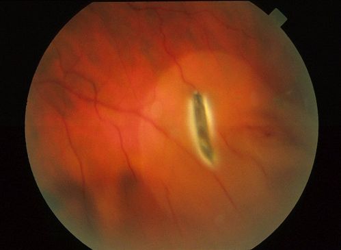
Retinal photoreceptors and RPE cells are especially susceptible to siderosis . Electroretinogram (ERG) changes in siderosis include an increased a-wave and normal b-wave during the very early phase of toxicity, with a diminishing b-wave amplitude later. Eventually, the ERG may become extinguished. Serial ERGs can be helpful for following eyes with small retained foreign bodies. If the b-wave amplitude decreases, removal of the foreign body generally is recommended.