
B SCAN ULTRASOUND (PER EYE)
B-scan ultrasonography is an important adjuvant for the clinical assessment of various ocular and orbital diseases.
The ultrasound uses high-frequency sound waves that travel through the eye. Reflections (echoes) of the sound waves form a picture of the structure of the eye.
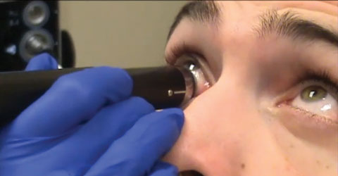
Your eye is numbed anesthetic eye drops. The ultrasound probe is placed against the front surface of the eye. The test takes about 15 minutes. You will be seated and you may be asked to look in many directions. The test is usually done with your eyes closed. A gel is placed on the skin of your eyelids. The B-scan probe is gently placed against your eyelids to do the test.
Your eye is numbed, so you shouldn’t have any discomfort. You may be asked to look in different directions to improve the ultrasound image or so it can view different areas of your eye.
B-scan is done to look at the inside part of the eye or the space behind the eye that can’t be seen directly. This may occur when you have cataracts or other conditions that make it hard for the doctor to see into the back of your eye. The test may help diagnose retinal detachment, tumors, or other disorders.